Home

semafor kontrast meme cholesteatoma diffusion mri tilki yeterlidir komplike

Role of diffusion-weighted MRI in the detection of cholesteatoma after  tympanoplasty - ScienceDirect
Role of diffusion-weighted MRI in the detection of cholesteatoma after tympanoplasty - ScienceDirect

The Utility of Diffusion-Weighted Imaging for Cholesteatoma Evaluation |  American Journal of Neuroradiology
The Utility of Diffusion-Weighted Imaging for Cholesteatoma Evaluation | American Journal of Neuroradiology

EPOS™
EPOS™

The Utility of Diffusion-Weighted Imaging for Cholesteatoma Evaluation |  American Journal of Neuroradiology
The Utility of Diffusion-Weighted Imaging for Cholesteatoma Evaluation | American Journal of Neuroradiology

Detection of Middle Ear Cholesteatoma by Diffusion-Weighted MR Imaging:  Multishot Echo-Planar Imaging Compared with Single-Shot Echo-Planar Imaging  | American Journal of Neuroradiology
Detection of Middle Ear Cholesteatoma by Diffusion-Weighted MR Imaging: Multishot Echo-Planar Imaging Compared with Single-Shot Echo-Planar Imaging | American Journal of Neuroradiology

The Utility of Diffusion-Weighted Imaging for Cholesteatoma Evaluation |  American Journal of Neuroradiology
The Utility of Diffusion-Weighted Imaging for Cholesteatoma Evaluation | American Journal of Neuroradiology

JPM | Free Full-Text | The Efficacy of DW and T1-W MRI Combined with CT in  the Preoperative Evaluation of Cholesteatoma
JPM | Free Full-Text | The Efficacy of DW and T1-W MRI Combined with CT in the Preoperative Evaluation of Cholesteatoma

Figure 1 from PROPELLER non-EPI DWI in the diagnosis of primary and  recurrent cholesteatoma . A pictorial review | Semantic Scholar
Figure 1 from PROPELLER non-EPI DWI in the diagnosis of primary and recurrent cholesteatoma . A pictorial review | Semantic Scholar

Contemporary Non–Echo-planar Diffusion-weighted Imaging of Middle Ear  Cholesteatomas | RadioGraphics
Contemporary Non–Echo-planar Diffusion-weighted Imaging of Middle Ear Cholesteatomas | RadioGraphics
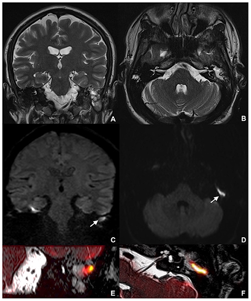
Frontiers | Combining Thin-Section Coronal and Axial Diffusion Weighted  Imaging: Good Practice in Middle Ear Cholesteatoma Neuroimaging
Frontiers | Combining Thin-Section Coronal and Axial Diffusion Weighted Imaging: Good Practice in Middle Ear Cholesteatoma Neuroimaging

A case of left middle ear cholesteatoma. MRI shows hypointense on... |  Download Scientific Diagram
A case of left middle ear cholesteatoma. MRI shows hypointense on... | Download Scientific Diagram

Cholesteatoma | Radiology Reference Article | Radiopaedia.org

The value of different diffusion-weighted magnetic resonance techniques in  the diagnosis of middle ear cholesteatoma. Is there still an indication for  echo-planar diffusion-weighted imaging?
The value of different diffusion-weighted magnetic resonance techniques in the diagnosis of middle ear cholesteatoma. Is there still an indication for echo-planar diffusion-weighted imaging?

Non-echoplanar diffusion weighted imaging in the detection of  post-operative middle ear cholesteatoma: navigating beyond the pitfalls to  find the pearl | Insights into Imaging | Full Text
Non-echoplanar diffusion weighted imaging in the detection of post-operative middle ear cholesteatoma: navigating beyond the pitfalls to find the pearl | Insights into Imaging | Full Text

The Utility of Diffusion-Weighted Imaging for Cholesteatoma Evaluation |  American Journal of Neuroradiology
The Utility of Diffusion-Weighted Imaging for Cholesteatoma Evaluation | American Journal of Neuroradiology
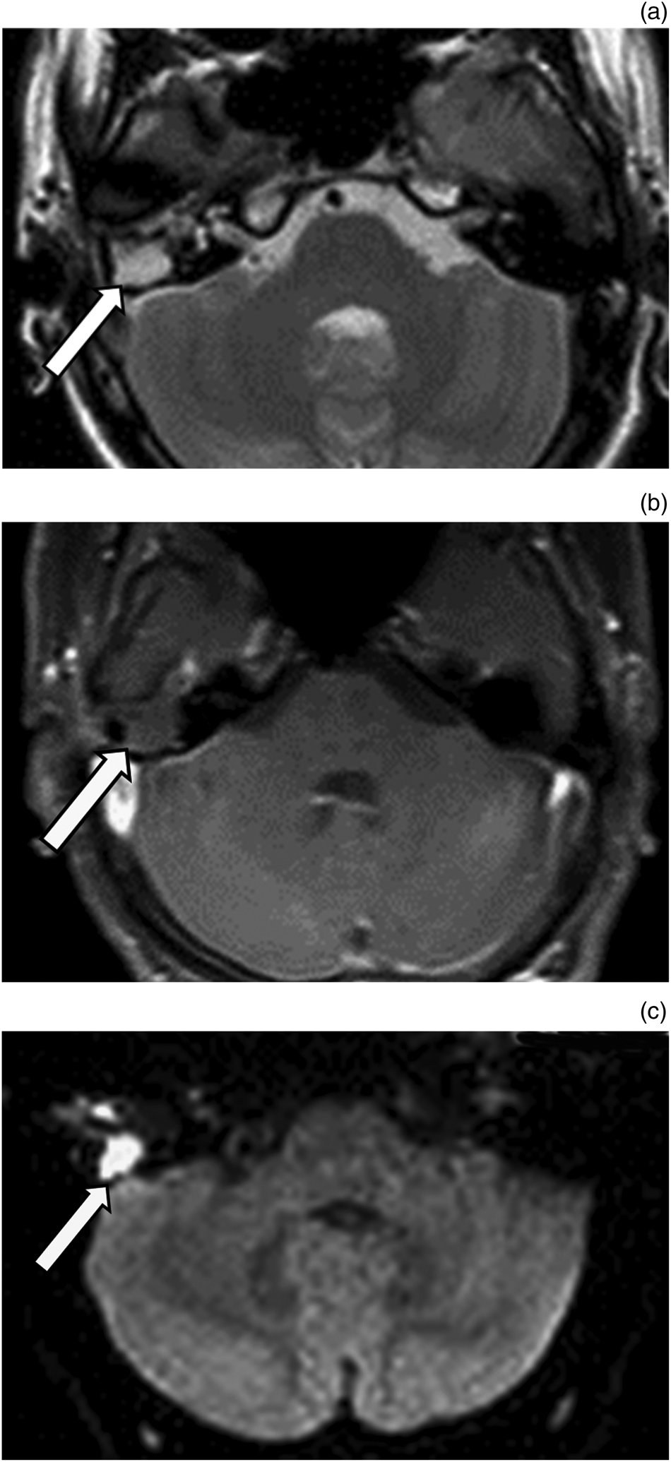
Reliability of diffusion-weighted magnetic resonance imaging in  differentiation of recurrent cholesteatoma and granulation tissue after  intact canal wall mastoidectomy | The Journal of Laryngology & Otology |  Cambridge Core
Reliability of diffusion-weighted magnetic resonance imaging in differentiation of recurrent cholesteatoma and granulation tissue after intact canal wall mastoidectomy | The Journal of Laryngology & Otology | Cambridge Core

Diffusion Weighted MR Imaging of Primary and Recurrent Middle Ear  Cholesteatoma: An Assessment by Readers with Different Expertise
Diffusion Weighted MR Imaging of Primary and Recurrent Middle Ear Cholesteatoma: An Assessment by Readers with Different Expertise

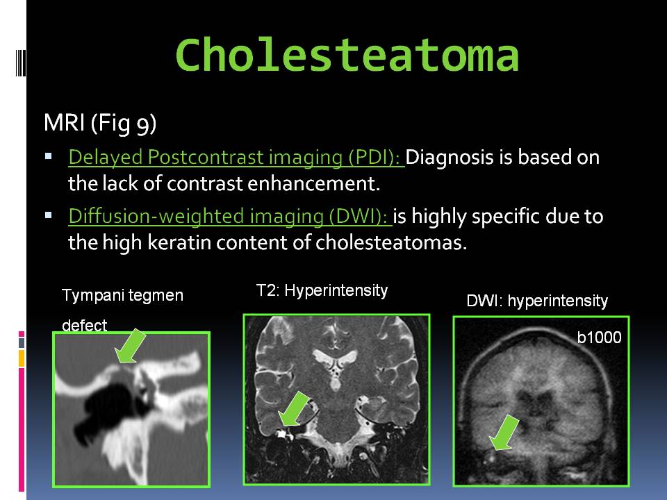
Role of diffusion-weighted MRI in the detection of cholesteatoma after  tympanoplasty - ScienceDirect
Role of diffusion-weighted MRI in the detection of cholesteatoma after tympanoplasty - ScienceDirect

Surgically confirmed cholesteatoma. a Non-EP DWI showing a high... |  Download Scientific Diagram
Surgically confirmed cholesteatoma. a Non-EP DWI showing a high... | Download Scientific Diagram

Cholesteatoma | Radiology Case | Radiopaedia.org